It's important to have regular eye tests so eye problems, such as glaucoma, can be diagnosed and treated as early as possible.
If you have glaucoma, it can take a long time before you realise you have a problem with your eyesight. This is because glaucoma usually damages the outer edge of the eye and works slowly inwards. You may not notice a problem until the glaucoma is near the centre of your eye.
You should have an eye test at least every two years or more frequently if advised by your optometrist (a healthcare professional who tests sight). For example, they may suggest you have more frequent eye tests if you have a close relative with glaucoma, such as a parent, brother or sister.
Tests for glaucoma
There are several glaucoma tests that are painless and quite quick. The tests will be carried out during the same appointment to ensure results are as accurate as possible.
These tests are explained below.
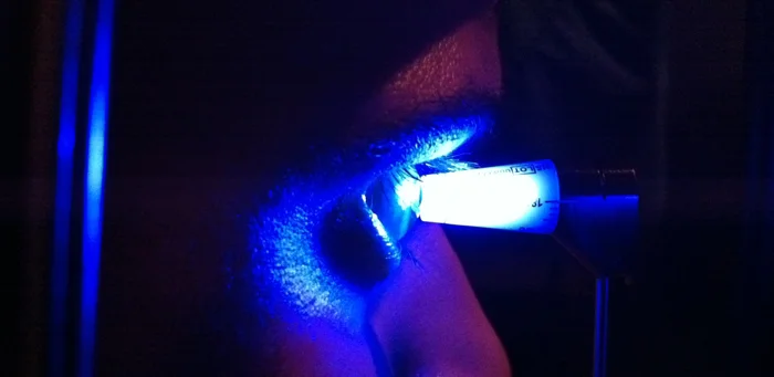
Eye pressure test (tonometry)
An eye pressure test (tonometry) uses an instrument called a tonometer to measure the pressure inside your eye.
A small amount of anaesthetic (painkilling medication) and dye is placed onto the transparent layer of tissue that covers the front of the eye (your cornea). A blue light from the head of the tonometer is held against your eye to measure the intraocular pressure.
Tonometry can diagnose ocular hypertension (OHT – raised pressure in the eye), which is a risk factor for chronic open-angle glaucoma.
Central corneal thickness
The thickness of your cornea will be measured because this is thought to affect how the intraocular pressure is interpreted.
Gonioscopy
Gonioscopy is an examination of the front outer edge of your eye, between the cornea and the iris (the coloured part of your eye). This is the area where the fluid should drain out of your eye.
A gonioscopy can help to determine whether this angle is open or closed (blocked).
Visual field test
A visual field test – sometimes called perimetry – checks for missing areas of vision. You will be shown a sequence of light spots and asked which ones you can see. Some dots will appear in your peripheral vision (around the sides of your eyeball), which is where glaucoma begins.
If you can't see the spots in your peripheral vision, it may indicate the glaucoma has damaged your vision.
Optic nerve assessment
Your optic nerve connects your eye to your brain. Your optometrist will use eye drops to enlarge your pupils. They will then examine your eyes using a slit lamp (a microscope with a very bright light) and assess whether your optic nerve has been damaged by the glaucoma.
The eye drops used to widen your pupils could affect your ability to drive. You should make alternative arrangements for getting home after your appointment.
OCT of Optic disc
Glaucomatous damage is often marked by retinal thinning in the zone surrounding the fovea and extending toward the optic nerve head. The SPECTRALIS Posterior Pole Asymmetry Analysis maps retinal thickness across the posterior pole and graphs asymmetry both between hemispheres and between eyes. Used with RNFL (Retinal nerve fiber layer) measurements using OCT (Optical Coherence Tomography), the Posterior Pole Asymmetry Analysis can provide a robust picture of glaucoma.
